Pages


Rabu, 26 Desember 2012

External compression headaches

3 komentar
External compression headaches

Definition:
External compression headaches

External compression headaches can occur when any head wear puts continuous pressure on your forehead or scalp.

Common culprits of external compression headaches include tight hats, helmets, headbands and goggles. These headaches are sometimes known by other names that are specific to the type of equipment causing your headache, such as "swim-goggle headache" or "football-helmet headache."
With external compression headaches, the solution is as obvious as the cause. Simply remove the head wear causing the pressure.

Symptoms:

The pain of external compression headaches is often described as constant pressure. It hurts most where the object is pressing on your head. As long as the head wear is in place, the pain may get progressively worse.

When to see a doctor
Because compression headaches go away when you remove whatever object is causing the pressure on your head, it's necessary to seek further medical attention only if your headache continues after you've removed the object.

Causes:

 External compression headaches are caused by any type of head wear that places pressure on the head — including tight hats, helmets, headbands and goggles


Treatments and drugs:

To end your headache, remove the head wear that's causing the pressure. Further treatment is rarely needed.

If you have a history of migraines, wearing certain head wear may trigger an attack that requires migraine medication for relief.
Read more...

Exercise-induced asthma

3 komentar
Exercise-induced asthma

Definition:
Exercise-induced asthma

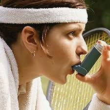
If you cough, wheeze or feel out of breath during or after exercise, it may be more than exertion causing your symptoms. You might have exercise-induced asthma. As with asthma triggered by other things, exercise-induced asthma symptoms occur when your airways tighten and produce extra mucus.

If you have exercise-induced asthma — also called exercise-induced bronchospasm (BRONG-ko-spaz-um) — physical exertion may be the only thing that triggers your symptoms. Or, exercise may be just one of several things that trigger your asthma. But having exercise-induced asthma doesn't mean you shouldn't exercise. Proper treatment of exercise-induced asthma and precautions can keep you active — whether you're strolling through the park or running a race.

Symptoms:

Exercise-induced asthma symptoms can include:
  • Coughing
  • Wheezing
  • Shortness of breath
  • Chest tightness or pain
  • Fatigue during exercise
  • Poor athletic performance
Exercise-induced asthma symptoms may start a few minutes after you begin exercising, and they may continue to worsen for another 10 minutes or so after you've finished a workout. It's possible to have symptoms both during and after exercise.

Feeling a little short of breath or fatigued when you work out is normal, especially if you aren't in great shape. But with exercise-induced asthma, these symptoms can be more severe.
For many people, exercise is just one of a few asthma triggers. Others can include pollen, pet dander and other airborne allergens.

When to see a doctor
See your doctor if you cough, wheeze, or have chest pain or tightness during or after exercise. Many people don't realize they have exercise-induced asthma because they think these are their body's normal responses to working out. Don't assume your symptoms are caused by being out of shape or short on endurance.

Seek immediate medical treatment if you have worsening symptoms. Severe asthma attacks can be life-threatening. Signs of an asthma attack that needs emergency treatment include:
  • Shortness of breath or wheezing that is quickly getting worse
  • No improvement even after using a rescue inhaler, such as an albuterol inhaler
  • Shortness of breath that continues even after you've recovered from your workout

Causes:

It isn't clear exactly what causes exercise-induced asthma, and why some people get it and others don't. In susceptible individuals, symptoms may be triggered by drying or cooling of the airways during heavy breathing.
Factors that can trigger or worsen exercise-induced asthma include:
  • Cold air
  • Dry air
  • Air pollution such as smoke or smog
  • High pollen counts
  • Having a respiratory infection such as a cold
  • Chemicals, such as chlorine in swimming pools
There's no particular exercise you must avoid when you have exercise-induced asthma, but activities that make you breathe hard are more likely to trigger symptoms. For example, aerobic exercise, such as running or playing basketball, hockey or soccer, is more likely to trigger symptoms than is
weightlifting, golfing or moderate-paced walking. Likewise, exercising in cold weather also can increase asthma symptoms because you're breathing in a lot of cold, dry air.

But don't let that discourage you. With proper treatment, you can do intense aerobic activities — and cold-weather workouts — without asthma symptoms slowing you down.

Complications:

Asthma of any kind — including exercise-induced asthma — may cause a number of complications. Proper treatment can help you avoid them. Possible asthma complications include:
  • Poor athletic performance
  • Permanent narrowing of the airways (bronchial tubes), which causes difficulty breathing
  • Emergency room visits and hospitalizations for severe asthma attacks

Treatments and drugs:

For many people, a few puffs from a quick-relief inhaler right before exercise is enough to control asthma symptoms. These bronchodilator (brong-koh-DIE-lay-tur) medications — albuterol, for example — quickly open the airways and can help control symptoms for several hours.

Quick-relief medications
Also called rescue medications, quick-relief medications are used as needed for rapid, short-term symptom relief during an asthma attack — or before exercise if your doctor recommends it. Types of quick-relief medications include:
  • Short-acting beta agonists. These inhaled bronchodilator medications include albuterol (ProAir HFA, Ventolin HFA, others), levalbuterol (Xopenex HFA) and pirbuterol (Maxair Autohaler). For most people, one to two puffs of albuterol or another inhaled short-acting beta agonist 10 to 15 minutes before exercise is enough to prevent symptoms for up to four hours. These inhaled bronchodilator medications can rapidly ease symptoms during an asthma attack. However, it is possible to develop a tolerance to these medications if they're used frequently.
  • Ipratropium (Atrovent). Your doctor might prescribe this inhaled medication for immediate relief of your symptoms. Like other bronchodilators, ipratropium relaxes the airways, making it easier to breathe. Ipratropium is mostly used for emphysema and chronic bronchitis, but it's sometimes used to treat asthma attacks.
However, some people also need to take additional medications to control asthma symptoms. You may need daily long-term control medications if you have frequent asthma symptoms when you're not exercising, or if using a medication before exercise doesn't keep your symptoms under control.

Long-term control medications
In most cases, these medications need to be taken every day. Types of long-term control medications include:
  • Inhaled corticosteroids. These medications include fluticasone (Flovent Diskus, Flovent HFA), budesonide (Pulmicort Flexhaler), mometasone (Asmanex Twisthaler), triamcinolone (Azmacort), flunisolide (Aerobid), beclomethasone (Qvar) and others. They are the most commonly prescribed type of long-term asthma medication. You may need to use these medications for several days to weeks before they reach their maximum benefit. Unlike oral corticosteroids, these corticosteroid medications have a relatively low risk of side effects and are generally safe for long-term use.
  • Leukotriene modifiers. These oral medications include montelukast (Singulair), zafirlukast (Accolate) and zileuton (Zyflo, Zyflo CR). They help prevent asthma symptoms for up to 24 hours. In rare cases, these medications have been linked to psychological reactions, such as agitation, aggression, hallucinations, depression and suicidal thinking. Seek medical advice right away for any unusual reaction.
  • Theophylline. This is a daily pill that helps keep the airways open (bronchodilator). Theophylline (Theo-24, Elixophyllin, others) relaxes the muscles around the airways to make breathing easier. It's not used as often now as in past years because more-effective medications are available.
  • Long-acting beta agonists (LABAs). These inhaled medications include salmeterol (Serevent Diskus) and formoterol (Foradil Aerolizer). Taken 30 minutes before exercise, long-acting beta agonists can prevent symptoms of exercise-induced asthma for up to 12 hours. However, they've been linked to severe asthma attacks. LABAs should always be taken only in combination with an inhaled corticosteroid.
  • Combination inhalers. Fluticasone and salmeterol (Advair Diskus), budesonide and formoterol (Symbicort), and mometasone and formoterol (Dulera) are examples of combination inhalers. These medications contain a LABA along with a corticosteroid. Like other LABA medications, these medications may increase your risk of a severe asthma attack and need to be used with caution.
Treatment for allergy-induced asthma
If your asthma is triggered or worsened by allergies, you may benefit from allergy treatment as well. Allergy treatments include:
  • Omalizumab (Xolair). This medication is specifically for people who have allergies and asthma. It reduces the immune system's reaction to allergy-causing substances, such as pollen, dust mites or pet dander. Xolair is delivered by injection.
  • Allergy medications. These include oral and nasal spray antihistamines and decongestants as well as corticosteroid, cromolyn and ipratropium nasal sprays.
  • Allergy shots (immunotherapy). Over time, immunotherapy injections gradually reduce your immune system reaction to specific allergens. This may help relieve your symptoms if certain allergens, such as pollen, trigger your asthma.
Don't rely only on quick-relief medications
If you've been prescribed long-term asthma control medications — such as inhaled corticosteroids — you shouldn't need to use your quick-relief inhaler more often than your doctor recommends. Keep a record of how many puffs you use each week. If you frequently need to use your quick-relief inhaler, see your doctor. You probably need to adjust your long-term control medication.
Read more...

Exercise headaches

2 komentar
Exercise headaches

Definition  :
Exercise headaches

Exercise headaches occur during or after sustained, strenuous exercise. Activities associated with exercise headaches include running, rowing, tennis, swimming and weightlifting.

Doctors divide exercise headaches into two categories. Primary exercise headaches are usually harmless, aren't connected to any underlying problems and can often be prevented with medication. Secondary exercise headaches, on the other hand, are caused by an underlying, often serious, problem within the brain — such as bleeding or a tumor — or outside the brain — such as coronary artery disease. Secondary exercise headaches may require emergency medical attention.

Symptoms:

Primary exercise headaches
These headaches:
  • Are usually described as throbbing
  • Occur during or after strenuous exercise
  • Affect both sides of the head in most cases
Secondary exercise headaches
These headaches may cause:
  • The same symptoms as primary exercise headaches
  • Vomiting
  • Loss of consciousness
  • Double vision
  • Neck rigidity
Primary exercise headaches typically last between five minutes and 48 hours, while secondary exercise headaches usually last at least a day and sometimes linger for several days or longer.
When to see a doctor
If you experience a headache during or after exercise, consult your doctor. Call your doctor right away if the headache begins abruptly or if it's your first headache of this type.

Causes:

Primary exercise headaches
The exact cause of primary exercise headaches is unknown. One theory is that strenuous exercise dilates blood vessels inside the skull.
Secondary exercise headaches
Secondary exercise headaches are caused by an underlying problem, such as:
  • Bleeding in the area between the brain and the thin membranes that cover the brain (subarachnoid hemorrhage)
  • Abnormalities in a blood vessel leading to or within the brain
  • Cancerous or noncancerous tumors
  • Obstruction of cerebrospinal fluid flow
  • Reduced blood flow in the arteries feeding the heart
  • Sinus infection

Treatments and drugs:

If no underlying structural or vascular problem is causing your exercise headaches, your doctor may recommend medications to take regularly to help prevent the headaches.
  • Indomethacin (Indocin), an anti-inflammatory drug, is commonly prescribed.
  • Propranolol, (Inderal, Inopran XL) a blood pressure medication, also is used to preventive exercise headaches.
If your exercise headaches are predictable, you may be able to take a medication an hour or two before a scheduled event, such as a tennis match or a hike at high altitude. Medications your doctor might prescribe include indomethacin and triptans (Imitrex, Relpax, others), which are commonly used to treat acute migraine headaches. If your exercise headaches are frequent or unpredictable, you might need to take the preventive medicine every day.
Read more...

Essential tremor

3 komentar
Essential tremor

Definition  :
Essential tremor

Essential tremor is a disorder of the nervous system that causes a rhythmic shaking. Essential tremor can affect almost any part of your body, but the trembling occurs most often in your hands — especially when you try to do simple tasks, such as drinking from a glass, tying shoelaces, writing or shaving. Essential tremor also may affect your head, voice, arms or legs.

Although usually not a dangerous condition, essential tremor worsens over time and can be severe in some people. It isn't caused by other diseases, although it's sometimes confused with Parkinson's disease. Essential tremor can occur at any age but is most common in older adults.

Symptoms:

Essential tremor signs and symptoms:
  • Begin gradually
  • Worsen with movement
  • Usually occur in the hands first, affecting one hand or both hands
  • Can include a "yes-yes" or "no-no" motion of the head
  • Are aggravated by emotional stress, fatigue, caffeine or extremes of temperature
Essential tremor vs. Parkinson's disease
Many people associate tremors with Parkinson's disease, but the two conditions differ in key ways:
  • When tremors occur. Essential tremor of the hands typically occurs when you use your hands. Tremors from Parkinson's are most prominent when your hands are at your sides or resting in your lap.
  • Associated conditions. Essential tremor doesn't cause other health problems, whereas Parkinson's is associated with a stooped posture, slow movement and a shuffling gait. However, people with essential tremor may sometimes develop other neurological signs and symptoms — such as an unsteady gait (ataxia).
  • Parts of body affected. Essential tremor can involve your hands, head, voice and legs. Tremors from Parkinson's typically affect your hands but not your head or voice.
Causes:

 About half of essential tremor cases appear to occur because of a genetic mutation. This is referred to as familial tremor. What causes essential tremor in people without a known genetic mutation isn't clear.

Complications:

Essential tremor is not life-threatening, but symptoms often worsen over time. If the tremors become severe, you may find it difficult to:
  • Hold a cup or glass without spilling
  • Eat normally
  • Put on makeup or shave
  • Talk, if your voice box or tongue is affected
  • Write — handwriting may become increasingly large, shaky and illegible

Treatments and drugs:

Some people with essential tremor may not require treatment if their symptoms are mild. But if your essential tremor is making it difficult to work or perform daily activities, you may want to discuss treatment options with your doctor.
Medications
  • Beta blockers. Normally used to treat high blood pressure, beta blockers — such as propranolol (Inderal), atenolol, metoprolol and nadolol — help relieve tremors in some people. They may not be an option if you also have asthma, diabetes or certain heart problems.
  • Anti-seizure medications. Epilepsy drugs — including primidone (Mysoline), gabapentin (Neurontin) and topiramate (Topamax) — may be effective in people who don't respond to beta blockers. The main side effects are drowsiness and flu-like symptoms, which usually disappear within a short time.
  • Tranquilizers. Doctors sometimes use drugs such as diazepam (Valium) and alprazolam (Xanax) to treat people whose tremors are made much worse by tension or anxiety. Side effects can include confusion and memory loss. Additionally, these medications should be used with caution because they can be habit-forming.
  • OnabotulinumtoxinA (Botox) injections. You're probably familiar with Botox as a treatment for facial wrinkles, but it can also be useful in treating some types of tremors, especially of the head and voice. Botox injections can improve problems for up to three months at a time. But if it's used to treat hand tremors, it can sometimes cause weakness in your fingers.
Therapy
Physical therapy exercises can sometimes reduce tremor and improve coordination and muscle control. Occupational therapists may suggest some of the following adaptive devices to reduce the effect of your tremors on your daily activities:
  • Heavier plates, glasses and utensils
  • Wrist weights
  • Wider writing implements
Surgery
Surgery may be an option for people whose tremors are severely disabling and who don't respond to medications. The type of surgery performed is known as deep brain stimulation.

Deep brain stimulation involves inserting a long, thin electrical probe into your thalamus — the portion of your brain responsible for causing your tremors. A wire from the probe is tunneled under your skin to your chest, where a pacemaker-like device has been inserted. This device transmits painless electrical pulses to interrupt signals from your thalamus that may be causing your tremors.

Side effects of surgery may include problems with motor control or speech, problems with balance and temporary or permanent cognitive impairment, such as learning difficulties, or problems with your vision. Deep brain stimulation, however, is very effective for severe essential tremor, and these side effects are rare.
Read more...

Essential thrombocythemia

1 komentar
Essential thrombocythemia

Definition  :
Essential thrombocythemia

Essential thrombocythemia is an uncommon disorder in which your body produces too many blood platelets (thrombocytes). It's also known as primary thrombocythemia (throm-boe-sigh-THEE-me-uh).

The most common symptoms of essential thrombocythemia include headache, lightheadedness, vision changes, and tingling, numbness or burning pain in the hands and feet. Essential thrombocythemia most often occurs in people over age 50 and is more common in women.

You may not need treatment for essential thrombocythemia if you don't have symptoms. If you have abnormal blood clotting or bleeding, however, medications can help you avoid potentially serious complications.

Symptoms:

Many people with essential thrombocythemia have no signs or symptoms. The first indication you have the disorder may be the development of a blood clot (thrombus). Although clots can develop anywhere in your body, with essential thrombocythemia, they occur most often in your brain, hands and feet.
Signs and symptoms depend on where the clot forms. They include:
  • Headache
  • Dizziness or lightheadedness
  • Chest pain
  • Weakness
  • Fainting
  • Temporary vision changes
  • Numbness or tingling of the hands and feet
  • Redness, throbbing and burning pain in the hands and feet (erythromelalgia)
  • Mildly enlarged spleen
Less commonly, essential thrombocythemia may cause bleeding, especially if your platelet count is extremely high (more than 1 million platelets per microliter of blood). Bleeding may take the form of:
  • Nosebleeds
  • Bruising
  • Bleeding from your mouth or gums
  • Bloody stool
A blood clot may cause a transient ischemic attack (TIA) — a temporary interruption of blood flow to part of the brain — or stroke. Signs and symptoms develop suddenly and include:
  • Weakness or numbness of your face, arm or leg, usually on one side of your body
  • Difficulty speaking or understanding speech (aphasia)
  • Blurred, double or decreased vision
When to see a doctor
If you have any signs or symptoms of abnormal blood clotting or bleeding, see your doctor.
If you develop signs or symptoms of a TIA or stroke, such as numbness or paralysis on one side of your body, seek medical attention immediately.


Causes:

Bone marrow — spongy tissue inside your bones — contains stem cells that can become red blood cells, white blood cells or platelets. Platelets travel through your blood vessels. They stick together to form clots that stop the bleeding when you damage a blood vessel, such as when you get a cut. A normal platelet count ranges from 150,000 to 450,000 platelets per microliter of blood.

If you have essential thrombocythemia, your bone marrow makes too many platelet-forming cells (megakaryocytes), which release too many platelets into your blood. The excess platelets may not function normally, leading to abnormal clotting or bleeding.

The exact cause of essential thrombocythemia and similar conditions, known as myeloproliferative neoplasms, isn't known. About half the people with the disorder have a mutation of the Janus kinase 2 (JAK2) gene. Other gene mutations also have been linked to essential thrombocythemia.
A high platelet count that's caused by an underlying condition such as an infection or iron deficiency is called reactive or secondary thrombocytosis.

Complications:

The abnormal blood clotting of essential thrombocythemia can lead to a variety of potentially serious complications, including:
  • Pregnancy complications. While many women who have thrombocythemia have normal, healthy pregnancies, be sure to have your doctor regularly monitor your condition. Uncontrolled thrombocythemia can cause miscarriage, premature delivery, high blood pressure (preeclampsia), early separation of the placenta from the uterine wall (placental abruption) and slow fetal growth. Your risk of complications may be reduced with regular checkups and medication.
  • Stroke. A clot that blocks blood flow to your brain can cause a stroke. If you develop signs and symptoms of a stroke, get immediate medical attention.
  • Heart attack. A clot that obstructs blood flow to your heart can cause a heart attack. If you develop signs and symptoms of a heart attack, such as pressure, fullness or a squeezing pain in the center of your chest lasting more than a few minutes; pain extending to your shoulder, arm, back, teeth or jaw; shortness of breath; and sweating or clammy skin, get immediate medical attention.
Essential thrombocythemia can also cause bleeding (hemorrhage) with significant blood loss. A small minority of people with essential thrombocythemia may later develop acute leukemia or myelofibrosis, both of which can be life-threatening:
  • Acute leukemia. Acute myelogenous leukemia is a type of blood and bone marrow cancer that progresses rapidly.
  • Myelofibrosis. This progressive bone marrow disorder results in bone marrow scarring, severe anemia, and enlargement of your liver and spleen.

Treatments and drugs:

Treatment of essential thrombocythemia depends on your risk of blood-clotting or bleeding episodes. If you're younger than 60, have had no signs or symptoms, and have no other risk factors for developing blood clots, such as smoking, you may simply need periodic medical checkups. If you're older than 60 and have had previous signs and symptoms of blood clots, your doctor likely will prescribe medication or a medical procedure to lower your platelet count. Your doctor may also recommend treatment if you have cardiovascular risk factors, such as high cholesterol, high blood pressure or diabetes.

Medication
If you're at risk of blood clots, your doctor may recommend low-dose aspirin, particularly if you're pregnant. Aspirin makes the platelets less sticky and your blood less likely to form clots. If you take aspirin during pregnancy, stop taking it at least one week before delivery to lower your risk of bleeding complications during delivery.

Drugs that reduce the platelet count and are commonly used to treat essential thrombocythemia include:
  • Hydroxyurea (Droxia, Hydrea). This drug suppresses bone marrow production of blood cells, including platelets. Also used to treat cancers, it's the most commonly prescribed platelet-lowering drug for essential thrombocythemia. It's often used in combination with low-dose aspirin. If you take hydroxyurea, your doctor will monitor your blood counts closely. There's some concern that long-term use may increase the risk of developing acute myelogenous leukemia.
  • Anagrelide (Agrylin). Unlike hydroxyurea, anagrelide isn't associated with an increased risk of leukemia. But it's not considered as effective as hydroxyurea. Side effects may include fluid retention, heart problems, headaches, dizziness, nausea and diarrhea.
  • Interferon alfa-2B (Intron A). Given by injection, this drug is less convenient to administer than hydroxyurea or anagrelide, may be more expensive and may cause less tolerable side effects. Side effects may include flu-like symptoms, confusion, nausea, depression, diarrhea, seizures, irritability and sleepiness.
Emergency plateletpheresis
Used only in emergencies, such as after a stroke or other dangerous blood clotting, a medical procedure known as plateletpheresis can be used to rapidly lower platelet count. During the procedure, an intravenous (IV) needle connected to a tube is inserted into one of your blood vessels. Your blood flows through the tube and into a device that removes platelets from your blood. The remaining portion of your blood (plasma) and your red cells are then returned to you through an IV line. The effect is temporary.
Read more...

Esophageal varices

1 komentar
Esophageal varices

Definition:
Esophageal varices

Esophageal varices are abnormal, enlarged veins in the lower part of the esophagus — the tube that connects the throat and stomach. Esophageal varices occur most often in people with serious liver diseases.

Esophageal varices develop when normal blood flow to your liver is slowed. The blood then backs up into nearby smaller blood vessels, such as those in your esophagus, causing the vessels to swell. Sometimes, esophageal varices can rupture, causing life-threatening bleeding.

A number of drugs and medical procedures are available to stop bleeding from esophageal varices. These same treatments can help prevent bleeding in people diagnosed with esophageal varices.


Symptoms:


Esophageal varices usually don't cause signs and symptoms unless they bleed. Signs and symptoms of bleeding esophageal varices include:
  • Vomiting blood
  • Black, tarry or bloody stools
  • Shock, in severe cases
When to see a doctor
Make an appointment with your doctor if you have any signs or symptoms that worry you. If you've been diagnosed with liver disease, ask your doctor about your risk of esophageal varices and how you may reduce your risk of these complications. Ask your doctor whether you should undergo an endoscopy procedure to check for esophageal varices.

If you've been diagnosed with esophageal varices, your doctor may instruct you to be vigilant for signs of bleeding. Bleeding esophageal varices are an emergency. Call 911 or your local emergency services right away if you experience bloody vomit or bloody stools.

Causes:

The enlarged veins of esophageal varices form when blood flow to your liver is slowed. Often the flow of blood is slowed by scar tissue in the liver caused by liver disease. When the blood to your liver is slowed, it begins to back up, leading to increased pressure within a major vein (portal vein) that carries blood to your liver. This pressure forces the blood into the nearby smaller veins, such as those in your esophagus. These fragile, thin-walled veins begin to balloon with the added blood. Sometimes the veins can rupture and bleed.

Liver diseases and other causes of esophageal varices
Esophageal varices are most often a complication of cirrhosis — irreversible scarring of the liver. Other diseases and conditions also can cause esophageal varices. Causes can include:
  • Severe liver scarring (cirrhosis). A number of liver diseases can result in cirrhosis, such as hepatitis infection, alcoholic liver disease and a bile duct disorder called primary biliary cirrhosis.
  • Blood clot (thrombosis). A blood clot in the portal vein or in a vein that feeds into the portal vein called the splenic vein can cause esophageal varices.
  • A parasitic infection. Schistosomiasis is a parasitic infection found in parts of Africa, South America, the Caribbean, the Middle East and Southeast Asia. The parasite can damage the liver, as well as the lungs, intestine and bladder.
  • A syndrome that causes blood to back up in your liver. Budd-Chiari syndrome is a rare condition that causes blood clots that can block the veins that carry blood out of your liver.

Complications:

Bleeding
The most serious complication of esophageal varices is bleeding. Once you have had a bleeding episode, your risk of another is greatly increased. In some cases, bleeding can cause the loss of so much blood volume that you go into shock. This can lead to death.

Treatments and drugs:


The primary aim in treating esophageal varices is to prevent bleeding. Bleeding esophageal varices are life-threatening. If bleeding occurs, treatments are available to try to stop the bleeding.

Treatments to prevent bleeding
Treatments to lower blood pressure in the portal vein may reduce the risk of bleeding esophageal varices. Treatments may include:
  • Medications to slow flow of blood in the portal vein. A type of blood pressure drug called a beta blocker may help reduce blood pressure in your portal vein, reducing the likelihood of bleeding. These medications include propranolol (Inderal, Innopran) and nadolol.
  • Using a scope to access your esophagus and treat varices. If your esophageal varices appear to have a very high risk of bleeding, your doctor may recommend some of the same treatments that are used to stop active bleeding. Treatments may involve using an endoscope to see inside your esophagus and inject a medication or tie off veins with an elastic band.
Treatments to stop bleeding
Bleeding varices are life-threatening, and immediate treatment is essential. Treatments used to stop bleeding include:
  • Using elastic bands to tie off bleeding veins. During variceal ligation, your doctor uses an endoscope to snare the varices and wrap them with an elastic band, which essentially "strangles" the veins so they can't bleed. Variceal ligation carries a small risk of complications, such as scarring of the esophagus.
  • Injecting a solution into bleeding veins. In a procedure called endoscopic injection therapy, the bleeding varices are injected with a solution that shrinks them. Complications can include perforation of the esophagus and scarring of the esophagus that can lead to trouble swallowing (dysphagia).
  • Medications to slow blood flow into the portal vein. Medications can slow the flow of blood from the internal organs to the portal vein, reducing the pressure in the vein. A drug called octreotide (Sandostatin) is often used in combination with endoscopic therapy to treat bleeding from esophageal varices. The drug is usually continued for five days after a bleeding episode.
  • Diverting blood flow away from the portal vein. A transjugular intrahepatic portosystemic shunt (TIPS) is a small tube that is placed between the portal vein and the hepatic vein, which carries blood from your liver back to your heart. By providing an additional path for blood, the shunt often can control bleeding from esophageal varices. But TIPS can cause a number of serious complications, including liver failure and mental confusion, which may develop when toxins that would normally be filtered by the liver are passed through the shunt directly into the bloodstream. TIPS is mainly used when all other treatments have failed or as a temporary measure in people awaiting a liver transplant.
  • Replacing the diseased liver with a healthy one. Liver transplant is an option for people with severe liver disease or those who experience recurrent bleeding of esophageal varices. Although liver transplantation is often successful, the number of people awaiting transplants far outnumbers the available organs.
Read more...

Esophageal spasms

1 komentar
Esophageal spasms

Definition:
Esophageal spasms

Esophageal spasms are painful muscle contractions that affect your esophagus, the hollow tube between your throat and your stomach. Esophageal spasms can feel like sudden, severe chest pain that lasts from a few minutes to hours.

Esophageal spasms usually occur only occasionally. But for some people, the muscle contractions are frequent and can prevent food and liquids from traveling through the esophagus. Esophageal spasms can lead to chronic pain and swallowing problems.

Treatment for occasional esophageal spasms may not be necessary. But if esophageal spasms interfere with your ability to eat or drink, treatments are available.


Symptoms:

Signs and symptoms of esophageal spasms can include:
  • Squeezing pain in your chest, often intense, which you might mistake for heart pain (angina)
  • Difficulty swallowing (dysphagia)
  • The feeling that an object is stuck in your throat (globus)
  • The return of food and liquids back up your esophagus (regurgitation)
When to see a doctor
The squeezing chest pain that esophageal spasms can cause can also be caused by a heart attack or angina. If you experience squeezing chest pain, especially if it is a new symptom, seek medical care right away to make sure it's not a heart problem.

Causes:

It's not clear what causes esophageal spasms.
A healthy esophagus normally moves food into your stomach through a series of coordinated muscle contractions (peristalsis). Esophageal spasms disrupt this process by making it difficult for the muscles in the walls of your lower esophagus to coordinate in order to move food to your stomach.

Types of esophageal spasms
Esophageal spasms may occur in two forms:
  • Diffuse esophageal spasms are occasional contractions in the esophageal muscles. This type of spasm is often accompanied by regurgitation of food or liquids.
  • Nutcracker esophagus is the term for painfully strong contractions in the esophageal muscles. Nutcracker esophagus is less likely to cause regurgitation of food and liquids.

Treatments and drugs:

If you experience esophageal spasms only occasionally, you may not need treatment. Your doctor may recommend avoiding food or situations that trigger your esophageal spasms.
If your esophageal spasms make it difficult to eat or drink, your doctor may recommend:
  • Managing any underlying conditions, such as heartburn, GERD, anxiety or depression. Treating these conditions may lessen the likelihood of esophageal spasm symptoms.
  • Medications to relax your swallowing muscles, which can reduce the severity of contractions.
  • Surgery is sometimes recommended to treat acid reflux that doesn't respond to medication, or to cut the muscle at the lower end of the esophagus, which can weaken esophageal contractions.
Read more...

Esophageal cancer

0 komentar
Esophageal cancer

Definition:
Esophageal cancer

Esophageal cancer is cancer that occurs in the esophagus — a long, hollow tube that runs from your throat to your stomach. Your esophagus carries food you swallow to your stomach to be digested.
Esophageal cancer usually begins in the cells that line the inside of the esophagus. Esophageal cancer can occur anywhere along the esophagus, but in people in the United States, it occurs most often in the lower portion of the esophagus. More men than women get esophageal cancer.

Esophageal cancer isn't common in the United States. In other areas of the world, such as Asia and parts of Africa, esophageal cancer is much more common.


Symptoms:

Signs and symptoms of esophageal cancer include:
  • Difficulty swallowing (dysphagia)
  • Weight loss without trying
  • Chest pain, pressure or burning
  • Fatigue
  • Frequent choking while eating
  • Indigestion or heartburn
  • Coughing or hoarseness
Early esophageal cancer typically causes no signs or symptoms.

When to see a doctor
Make an appointment with your doctor if you have any persistent signs and symptoms that worry you.
If you've been diagnosed with Barrett's esophagus, a precancerous condition that increases your risk of esophageal cancer caused by chronic acid reflux, ask your doctor what signs and symptoms to watch for that may signal that your condition is worsening.

Screening for esophageal cancer isn't done routinely because of a lack of an easily identifiable high-risk group and the possible risks associated with endoscopy. If you have Barrett's esophagus, discuss the pros and cons of screening with your doctor.


Causes:

It's not clear what causes esophageal cancer. Esophageal cancer occurs when cells in your esophagus develop errors (mutations) in their DNA. The errors make cells grow and divide out of control. The accumulating abnormal cells form a tumor in the esophagus that can grow to invade nearby structures and spread to other parts of the body.

Types of esophageal cancer
Esophageal cancer is classified according to the type of cells that are involved. The type of esophageal cancer you have helps determine your treatment options. Types of esophageal cancer include:
  • Adenocarcinoma. Adenocarcinoma begins in the cells of mucus-secreting glands in the esophagus. Adenocarcinoma occurs most often in the lower portion of the esophagus. Adenocarcinoma is the most common form of esophageal cancer in the United States, and it affects primarily white men.
  • Squamous cell carcinoma. The squamous cells are flat, thin cells that line the surface of the esophagus. Squamous cell carcinoma occurs most often in the middle of the esophagus. Squamous cell carcinoma is the most prevalent esophageal cancer worldwide.
  • Other rare types. Rare forms of esophageal cancer include choriocarcinoma, lymphoma, melanoma, sarcoma and small cell cancer.

Complications:

As esophageal cancer advances, it can cause complications, such as:
  • Obstruction of the esophagus. Cancer may make it difficult or impossible for food and liquid to pass through your esophagus.
  • Cancer pain. Advanced esophageal cancer can cause pain.
  • Bleeding in the esophagus. Esophageal cancer can cause bleeding. Though bleeding is usually gradual, it can be sudden and severe at times.
  • Severe weight loss. Esophageal cancer can make it difficult and painful to eat and drink, resulting in weight loss.
  • Coughing. Esophageal cancer can erode your esophagus and create a hole into your windpipe (trachea). Known as a tracheoesophageal fistula, this hole can cause severe and sudden coughing whenever you swallow.

Treatments and drugs:

What treatments you receive for esophageal cancer are based on the type of cells involved in your cancer, your cancer's stage, your overall health and your preferences for treatment.

Surgery
Surgery to remove the cancer can be used alone or in combination with other treatments. Operations used to treat esophageal cancer include:
  • Surgery to remove very small tumors. If your cancer is very small, confined to the superficial layers of your esophagus and hasn't spread, your surgeon may recommend removing the cancer and margin of healthy tissue that surrounds it. Surgery for very early-stage cancers can be done using an endoscope passed down your throat and into your esophagus.
  • Surgery to remove a portion of the esophagus (esophagectomy). Your surgeon removes the portion of your esophagus that contains the tumor and nearby lymph nodes. The remaining esophagus is reconnected to your stomach. Usually this is done by pulling the stomach up to meet the remaining esophagus. In some situations, a portion of the colon is used to replace the missing section of esophagus.
  • Surgery to remove part of your esophagus and the upper portion of your stomach (esophagogastrectomy). Your surgeon removes part of your esophagus, nearby lymph nodes and the upper part of your stomach. The remainder of your stomach is then pulled up and reattached to your esophagus. If necessary, part of your colon is used to help join the two.
Esophageal cancer surgery carries a risk of serious complications, such as infection, bleeding and leakage from the area where the remaining esophagus is reattached. Surgery to remove your esophagus can be performed as an open procedure using large incisions or with special surgical tools inserted through several small incisions in your skin (laparoscopically). How your surgery is performed depends on your situation and your surgeon's experience and preferences.

Surgery for supportive care
Besides treating the disease, surgery can help relieve symptoms or allow you to eat.
  • Relieving esophageal obstruction. A number of treatments are available to relieve esophageal obstruction. One option includes using an endoscope and special tools to widen the esophagus and place a metal tube (stent) to hold the esophagus open. Other options include surgery, radiation therapy, chemotherapy, laser therapy and photodynamic therapy.
  • Providing nutrition. A surgeon inserts a feeding tube (percutaneous gastronomy) so you can receive nutrition directly into your stomach or intestine. This is usually temporary until the surgical site heals or until you're finished with chemotherapy and radiation therapy.
Chemotherapy
Chemotherapy is drug treatment that uses chemicals to kill cancer cells. Chemotherapy drugs are typically used before (neoadjuvant) or after (adjuvant) surgery in people with esophageal cancer. Chemotherapy can also be combined with radiation therapy. In people with advanced cancer that has spread beyond the esophagus, chemotherapy may be used alone to help relieve signs and symptoms caused by the cancer.

The chemotherapy side effects you experience depend on which chemotherapy drugs you receive.

Radiation therapy
Radiation therapy uses high-powered energy beams to kill cancer cells. Radiation can come from a machine outside your body that aims the beams at your cancer (external beam radiation). Or radiation can be placed inside your body near the cancer (brachytherapy).

Radiation therapy is most often combined with chemotherapy in people with esophageal cancer. It can be used before or after surgery. Radiation therapy is also used to relieve complications of advanced esophageal cancer, such as when a tumor grows large enough to stop food from passing to your stomach.

Side effects of radiation to the esophagus include sunburn-like skin reactions, painful or difficult swallowing, and accidental damage to nearby organs, such as the lungs and heart.

Combined chemotherapy and radiation
Combining chemotherapy and radiation therapy may enhance the effectiveness of each treatment. Combined chemotherapy and radiation may be the only treatment you receive, or combined therapy can be used before surgery. But combining chemotherapy and radiation treatments increases the likelihood and severity of side effects.

Clinical trials
Clinical trials are research studies testing the newest cancer treatments and new ways of using existing cancer treatments. Clinical trials give you a chance to try the latest in cancer treatment, but they can't guarantee a cure. Ask your doctor if you're eligible to enroll in a clinical trial. Together you can discuss the potential benefits and risks.
Read more...

Epilepsy

3 komentar
Epilepsy

Definition:
Epilepsy

Epilepsy is a disorder that results from the surges in electrical signals inside the brain, causing recurring seizures. Seizure symptoms vary. Some people with epilepsy simply stare blankly for a few seconds during a seizure, while others have full-fledged convulsions.

About 2 in 100 people in the United States will experience an unprovoked seizure once in life. However, a solitary seizure doesn't mean you have epilepsy. At least two unprovoked seizures are generally required for an epilepsy diagnosis.

Even mild seizures may require treatment because they can be dangerous during activities such as driving or swimming. Treatment — which generally includes medications and sometimes surgery — usually eliminates or reduces the frequency and intensity of seizures. Many children with epilepsy even outgrow the condition with age.

Symptoms:

Because epilepsy is caused by abnormal activity in brain cells, seizures can affect any process your brain coordinates. A seizure can produce:
  • Temporary confusion
  • A staring spell
  • Uncontrollable jerking movements of the arms and legs
  • Loss of consciousness or awareness
Symptoms vary depending on the type of seizure. In most cases, a person with epilepsy will tend to have the same type of seizure each time, so the symptoms will be similar from episode to episode.
Doctors generally classify seizures as either focal or generalized, based on how the abnormal brain activity begins.

Focal seizures
When seizures appear to result from abnormal activity in just one part of the brain, they're called focal or partial seizures. These seizures fall into two categories.
  • Simple focal seizures. These seizures don't result in loss of consciousness. They may alter emotions or change the way things look, smell, feel, taste or sound. They may also result in involuntary jerking of part of the body, such as an arm or leg, and spontaneous sensory symptoms such as tingling, vertigo and flashing lights.
  • Complex focal seizures. These seizures alter consciousness or awareness, causing you to lose awareness for a period of time. Complex focal seizures often result in staring and nonpurposeful movements — such as hand rubbing, chewing, swallowing or walking in circles.
Generalized seizures
Seizures that seem to involve all of the brain are called generalized seizures. Six types of generalized seizures exist.
  • Absence seizures (also called petit mal). These seizures are characterized by staring and subtle body movement, and can cause a brief loss of awareness.
  • Tonic seizures. These seizures cause stiffening of the muscles, generally those in your back, arms and legs and may cause you to fall to the ground.
  • Clonic seizures. These types of seizures are associated with rhythmic, jerking muscle contractions, usually affecting the arms, neck and face.
  • Myoclonic seizures. These seizures usually appear as sudden brief jerks or twitches of your arms and legs.
  • Atonic seizures. Also known as drop attacks, these seizures cause you to lose normal muscle tone and suddenly collapse or fall down.
  • Tonic-clonic seizures (also called grand mal). The most intense of all types of seizures, these are characterized by a loss of consciousness, body stiffening and shaking, and sometimes loss of bladder control or biting your tongue.
When to see a doctor
Seek immediate medical help if any of the following occurs:
  • The seizure lasts more than five minutes.
  • Breathing or consciousness does not return after the seizure stops.
  • A second seizure follows immediately.
  • You have a high fever.
  • You're experiencing heat exhaustion.
  • You're pregnant.
  • You have diabetes.
  • You've injured yourself during the seizure.
If you experience a seizure for the first time, seek medical advice.


Causes:

Epilepsy has no identifiable cause in about half of those who have the condition. In the other half, the condition may be traced to various factors.
  • Genetic influence. Some types of epilepsy, which are categorized by your type of seizure, run in families, making it likely that there's a genetic influence. Researchers have linked some types of epilepsy to specific genes, though it's estimated that up to 500 genes could be tied to the condition. For most people, genes are only part of the cause, perhaps by making a person more susceptible to environmental conditions that trigger seizures.
  • Head trauma sustained during a car accident or other traumatic injury can cause epilepsy.
  • Medical disorders. Events like strokes or heart attacks that result in damage to the brain also can cause epilepsy. Stroke is responsible for up to one-half of epilepsy cases in those over age 35.
  • Dementia is a leading cause of epilepsy among older adults.
  • Diseases such as meningitis, AIDS and viral encephalitis can cause epilepsy.
  • Prenatal injury. Before birth, babies are susceptible to brain damage caused by an infection in the mother, poor nutrition or oxygen deficiencies. This can lead to cerebral palsy in the child. About 20 percent of seizures in children are associated with cerebral palsy or other neurological abnormalities.
  • Developmental disorders. Epilepsy can sometimes be associated with other developmental disorders, such as autism and Down syndrome.

Complications:

Having a seizure at certain times can lead to circumstances that are dangerous to yourself or others.
  • Falling. If you fall during a seizure, you can injure your head or break a bone.
  • Drowning. If you have epilepsy, you're 13 times more likely to drown while swimming or bathing than is the rest of the population because of the possibility of having a seizure while in the water.
  • Car accidents. A seizure that causes either loss of awareness or control can be dangerous if you're driving a car or operating other equipment. Many states have driver's-licensing restrictions related to your ability to control seizures and impose a minimum amount of time that you've been seizure-free — ranging from three months to two years — before you're allowed to drive.
  • Pregnancy complications. Seizures during pregnancy pose dangers to both mother and baby, and certain anti-epileptic medications increase the risk of birth defects. If you have epilepsy and you're considering becoming pregnant, talk to your doctor as you plan your pregnancy. Most women with epilepsy can become pregnant and have a healthy baby. You'll need to be carefully monitored throughout pregnancy, and medications may need to be adjusted. It's very important that you work with your doctor to plan your pregnancy.
  • Emotional health issues. People with epilepsy are more prone to have psychological problems, especially depression, anxiety and, in extreme cases, suicide. This could be due to difficulties dealing with the condition itself as well as medication side effects.
Other life-threatening complications from epilepsy are uncommon, but do occur.
  • Status epilepticus. This condition occurs if you're in a state of continuous seizure activity lasting more than five minutes, or you have frequent recurrent seizures without regaining full consciousness in between them. People with status epilepticus have an increased risk of permanent brain damage and death.
  • Sudden unexplained death in epilepsy (SUDEP). People with poorly controlled epilepsy also have a small risk of sudden unexplained death. Overall, less than 1 in 1,000 people with epilepsy die of SUDEP, but it's more common among people whose seizures aren't controlled by treatment. The risk of SUDEP is particularly elevated when generalized tonic-clonic seizures are frequent, and the risk over a one-year period could be as high as approximately 1 in a hundred people.

Treatments and drugs:

Doctors generally start by treating epilepsy with medication. If that doesn't work, they may propose surgery or another type of treatment.

Medication
Most people with epilepsy can become seizure-free by using a single anti-epileptic drug. Others can decrease the frequency and intensity of their seizures. More than half the children with medication-controlled epilepsy can eventually stop medications and live a seizure-free life. Many adults also can discontinue medication after two or more years without seizures.

Finding the right medication and dosage can be complex. Your doctor likely will first prescribe a single drug at a relatively low dosage and may increase the dosage gradually until your seizures are well controlled.

All anti-seizure medications have some side effects. Mild side effects include:
  • Fatigue
  • Dizziness
  • Weight gain
  • Loss of bone density
  • Skin rashes
  • Loss of coordination
  • Speech problems
More severe but rare side effects include:
  • Depression
  • Suicidal thoughts and behaviors
  • Severe rash
  • Inflammation of certain organs, such as your pancreas or liver
To achieve the best seizure control possible with medication:
  • Take medications exactly as prescribed.
  • Always call your doctor before switching to a generic version of your medication or taking other prescription medications, over-the-counter drugs or herbal remedies.
  • Never stop taking your medication without talking to your doctor.
  • Notify your doctor immediately if you notice new or increased feelings of depression, suicidal thoughts or unusual changes in your mood or behaviors.
At least half of all people newly diagnosed with epilepsy will become seizure-free with their first medication. If anti-epileptic medications don't provide satisfactory results, your doctor may suggest surgery or other therapies.

Surgery
Surgery is most commonly done when tests show that your seizures originate in a small, well-defined area of your brain that doesn't interfere with vital functions like speech, language or hearing. In these types of surgeries, your doctor removes the area of the brain that's causing the seizures.
If your seizures originate in a part of your brain that can't be removed, your doctor may recommend a different sort of surgery in which surgeons make a series of cuts in your brain. These cuts are designed to prevent seizures from spreading to other parts of the brain.

Although many people continue to need some medication to help prevent seizures after successful surgery, you may be able to take fewer drugs and reduce your dosages. In a small number of cases, surgery for epilepsy can cause complications such as permanently altering your cognitive abilities. Talk to your surgeon about his or her experience, success rates and complication rates with the procedure you're considering.

Therapies
  • Vagus nerve stimulation. This therapy involves a device called a vagus nerve stimulator that's implanted underneath the skin of your chest like a heart pacemaker. Wires from the stimulator are wrapped around the vagus nerve in your neck. The battery-powered device delivers short bursts of electrical energy to the brain through the vagus nerve. It's not clear how this inhibits seizures, but the device can reduce seizures by 20 to 40 percent and completely control seizures in about 5 percent of people. Most people still need to take anti-epileptic medication. Side effects of vagus nerve stimulation include hoarseness, throat pain, coughing, shortness of breath, tingling and muscle pain.
  • Ketogenic diet. Some children with epilepsy have been able to reduce their seizures by maintaining a strict diet that's high in fats and low in carbohydrates. This diet, called a ketogenic diet, causes the body to break down fats instead of carbohydrates for energy. Some children can go off the ketogenic diet after a few years and remain seizure-free.

    Consult a doctor if you or your child is considering a ketogenic diet. It's important to make sure that a child doesn't become malnourished when taking the diet. Side effects of a ketogenic diet may include dehydration, constipation, slowed growth because of nutritional deficiencies, and buildup of uric acid in the blood, which can cause kidney stones. These side effects are uncommon if use of the diet is properly and medically supervised.
Read more...

Epiglottitis

1 komentar
Epiglottitis
Epiglottitis

Definition:

Epiglottitis is a potentially life-threatening condition that occurs when the epiglottis — a small cartilage "lid" that covers your windpipe — swells, blocking the flow of air into your lungs.

A number of factors can cause the epiglottis to swell — burns from hot liquids, direct injury to your throat and various infections. The most common cause of epiglottitis in children in the past was infection with Haemophilus influenzae type b (Hib), the same bacterium that causes pneumonia, meningitis and infections in the bloodstream. Epiglottitis can occur at any age.

Routine Hib vaccination for infants has made epiglottitis rare, but epiglottitis remains a concern. If you suspect that you or someone in your family has epiglottitis, seek emergency help immediately. Prompt treatment can prevent life-threatening complications.

Symptoms:

Symptoms in children
In children, signs and symptoms of epiglottitis may develop within a matter of hours, including:
  • Fever
  • Severe sore throat
  • Abnormal, high-pitched sound when breathing in (stridor)
  • Difficult and painful swallowing
  • Drooling
  • Anxious, restless behavior
  • Greater comfort when sitting up or leaning forward
Symptoms in adults
For adults, signs and symptoms may develop more slowly, over days rather than hours. Signs and symptoms may include:
  • Severe sore throat
  • Fever
  • A muffled or hoarse voice
  • Abnormal, high-pitched sound when breathing in (stridor)
  • Difficulty breathing
  • Difficulty swallowing
When to see a doctor
Epiglottitis is a medical emergency. If you or someone you know suddenly has trouble breathing and swallowing, call your local emergency number or go to the nearest hospital emergency department. Try to keep the person quiet and upright, because this position may make it easier to breathe. Don't try to examine the person's throat yourself. This can make matters worse.


Causes:

Epiglottitis is caused by an infection or an injury.
Infection
A common cause of swelling and inflammation of the epiglottis and surrounding tissues is infection with Haemophilus influenzae type b (Hib) bacteria. Hib is responsible for a number of serious conditions, the most common of which is meningitis.

Hib spreads through infected droplets coughed or sneezed into the air. It's possible to harbor Hib in your nose and throat without becoming sick — though you still have the potential to spread the bacteria to others.

Other bacteria and viruses also can cause inflammation of the epiglottis, including:
  • Streptococcus pneumoniae (pneumococcus), another bacterium that can cause meningitis, pneumonia, ear infections and blood infection (septicemia)
  • Streptococcus A, B and C, a group of bacteria that also can cause diseases ranging from strep throat to blood infections
Injury
Physical injury, such as a direct blow to the throat, can cause epiglottitis. So can burns from drinking very hot liquids.
You also may develop signs and symptoms similar to those of epiglottitis if you:
  • Swallow a chemical that burns your throat
  • Swallow a foreign object
  • Smoke drugs, such as crack cocaine

Complications:

Epiglottis can cause a number of complications, including:
  • Respiratory failure. The epiglottis is a small, movable "lid" just above the larynx that prevents food and drink from entering your windpipe. But if the epiglottis becomes swollen — either from infection or from injury — the airway narrows and may become completely blocked. This can lead to respiratory failure — a life-threatening condition in which the level of oxygen in the blood drops dangerously low or the level of carbon dioxide becomes excessively high.
  • Spreading infection. Sometimes the bacteria that cause epiglottitis cause infections elsewhere in the body, such as pneumonia, meningitis or a blood infection (sepsis).

Treatments and drugs:

Treatment of epiglottis involves first making sure you or your child can breathe, and then treating any identified infection.

Helping you breathe
The first priority in treating epiglottitis is ensuring that you or your child is receiving enough air. This may mean:
  • Wearing a mask. The mask delivers oxygen to the lungs.
  • Having a breathing tube placed into the windpipe through the nose or mouth (intubation). The tube must remain in place until the swelling in your or your child's throat has decreased — sometimes for several days.
  • Inserting a needle into the trachea (needle tracheostomy). In extreme cases or if more conservative measures fail, the doctor may need to create an emergency airway by inserting a needle directly into an area of cartilage in your or your child's trachea. This procedure allows air into your lungs while bypassing the larynx.
Treating infection
If your epiglottitis is related to an infection, intravenous antibiotics will be given once you or your child is getting enough air.
  • Broad-spectrum antibiotic. Because of the need for quick treatment, rather than wait for the results of the blood and tissue cultures, you or your child is likely to be treated with a broad-spectrum drug.
  • More targeted antibiotic. The drug may be changed later, depending on what's causing the epiglottitis.
Read more...

Epididymitis

13 komentar
Epididymitis

Definition:
Epididymitis

Epididymitis is an inflammation of the coiled tube (epididymis) at the back of the testicle that stores and carries sperm. Pain and swelling are the most common signs and symptoms of epididymitis. Males of any age can get epididymitis, but it's most common in men between the ages of 14 and 35.

Epididymitis is most often caused by a bacterial infection or by a sexually transmitted infection (STI), such as gonorrhea or chlamydia. In some cases, the testicle also may become inflamed — a condition called epididymo-orchitis.


Symptoms:

Epididymitis symptoms depend on the cause. They can include:
  • A tender, swollen, red or warm scrotum
  • Testicle pain and tenderness, usually on one side — the pain may get worse when you have a bowel movement
  • Painful urination or an urgent or frequent need to urinate
  • Painful intercourse or ejaculation
  • Chills and a fever
  • A lump on the testicle
  • Enlarged lymph nodes in the groin (inguinal nodes)
  • Pain or discomfort in the lower abdomen or pelvic area
  • Discharge from the penis
  • Blood in the semen
Chronic epididymitis
Signs and symptoms of epididymitis usually develop over a day or two and get better with treatment. In some cases, epididymitis may not clear up completely or may recur. This is known as chronic epididymitis. Symptoms of chronic epididymitis may come on gradually. Sometimes the cause of chronic epididymitis is not identified.

When to see a doctor
Never ignore scrotal pain or swelling. A number of conditions can cause testicular pain, and some of the conditions require immediate treatment. Some conditions that cause scrotal pain or swelling can get worse or cause permanent damage if not treated promptly.

Testicular torsion, caused by a twisted spermatic cord, can cause pain similar to that caused by epididymitis and requires emergency treatment because it blocks blood flow to the testicle.
See a doctor if you have discharge from your penis or pain when you urinate


Causes:


Epididymitis has a number of causes, including:
  • Sexually transmitted infections (STIs). STIs, particularly gonorrhea and chlamydia, are the most common cause of epididymitis in young, sexually active men.
  • Other infections. Boys, older men and homosexual men are more likely to have epididymitis caused by a nonsexually transmitted bacterial infection. For men and boys who've had urinary tract infections or prostate infections, bacteria may spread from the infected site to the epididymis. Rarely, epididymitis is caused by a fungal infection.
  • The heart medication amiodarone (Pacerone). In some cases, this anti-arrhythmic medication causes inflammation of the epididymis. Epididymitis caused by amiodarone is treated by temporarily discontinuing the drug or reducing the dose.
  • Tuberculosis. In some cases, tuberculosis can cause epididymitis.
  • Urine in the epididymis. Known as chemical epididymitis, this occurs when urine flows backward into the epididymis. It may occur with heavy lifting or straining.
Epididymitis:

Epididymitis may eventually cause:
  • Scrotal abscess, when infected tissue fills with pus
  • Chronic epididymitis, which can occur when untreated acute epididymitis leads to recurrent episodes
  • Shrinkage of the affected testicle (atrophy)
  • Reduced fertility, but this is rare
If the condition spreads from your epididymis to your testicle, the resulting condition is known as epididymo-orchitis. Signs, symptoms and treatment options are basically the same as they are for epididymitis.


Treatments and drugs:

Epididymitis caused by a sexually transmitted infection (STI) or other infection is treated with antibiotic medications. Your sexual partner will also need treatment. Make sure your doctor is aware of any other medications you're taking or any allergies you have. This information, as well as determining what type of infection you have, will help your doctor select the best treatment.

Be sure to take the entire course of antibiotics prescribed by your doctor, even though you may feel better in one to three days after you start treatment. If you're not feeling better in that time, contact your doctor.

When you've finished your medication, it's a good idea to return to your doctor for a follow-up visit to be sure that the infection has cleared up. If it hasn't, your doctor may try another antibiotic. If the infection still doesn't clear, your doctor may do further tests to determine whether your epididymitis is caused by something other than a bacterial infection or an STI.

If a pocket of pus (abscess) has formed, it may need to be drained, and in some cases part or all of the epididymis needs to be removed surgically (epididymectomy). Surgery may also be considered if epididymitis is due to underlying physical defects, but many people continue to have scrotal pain following epididymectomy.
Read more...

Epidermolysis bullosa

2 komentar
Epidermolysis bullosa

Definition  :
Epidermolysis bullosa

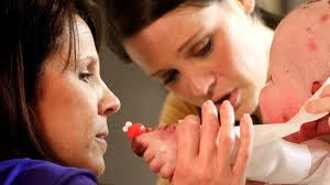
Epidermolysis bullosa (ep-ih-dur-MOL-uh-sis buhl-LO-sah) is a group of skin conditions whose hallmark is blistering in response to minor injury, heat, or friction from rubbing, scratching or adhesive tape. Four main types of epidermolysis bullosa exist, with numerous subtypes. Most are inherited.

Most types of epidermolysis bullosa initially affect infants and young children, although some people with mild forms of the condition don't develop signs and symptoms until adolescence or early adulthood. Mild forms of epidermolysis bullosa may improve with age, but severe forms may cause serious complications and can be fatal.

There's currently no cure for epidermolysis bullosa. For now, treatment focuses on addressing the symptoms, including pain prevention, wound prevention, infection and severe itching that occurs with continuous wound healing.

Symptoms:


The primary indication of epidermolysis bullosa is the eruption of fluid-filled blisters (bullae) on the skin, most commonly on the hands and feet in response to friction. Blisters of epidermolysis bullosa typically develop in various areas, depending on the type. In mild cases, blisters heal without scarring.
Signs and symptoms of epidermolysis bullosa may include:
  • Blistering of your skin — how widespread and severe depends on the type
  • Deformity or loss of fingernails and toenails
  • Internal blistering, including on the throat, esophagus, upper airway, stomach, intestines and urinary tract
  • Skin thickening on palms and soles of the feet (hyperkeratosis)
  • Scalp blistering, scarring and hair loss (scarring alopecia)
  • Thin-appearing skin (atrophic scarring)
  • Tiny white skin bumps or pimples (milia)
  • Dental abnormalities, such as tooth decay from poorly formed tooth enamel
  • Excessive sweating
  • Difficulty swallowing (dysphagia)
When to see a doctor
Contact your doctor promptly if you or your child develops blisters, particularly if there's no apparent reason for them.

In some cases of epidermolysis bullosa, blistering may not appear until a toddler first begins to walk, or until an older child begins new physical activities that trigger more intense friction on the feet.

Call your doctor immediately if you or your child experiences problems swallowing or breathing.
Also seek immediate care if you or your child has been diagnosed with epidermolysis bullosa and develops signs of an infection around an open area of skin, including:
  • Redness, heat or pain
  • Pus or a yellow discharge
  • Crusting
  • A red line or streak under the skin, spreading outward from the wound
  • A wound that does not heal
  • Fever or chills
Blisters can lead to infection and deformity. Your doctor can show you how to care for them properly and advise you on ways to prevent them


Causes:


In most cases, epidermolysis bullosa is inherited. Researchers have identified more than 10 genes involved with skin formation that, if defective, may cause a type of epidermolysis bullosa. It's also possible to develop epidermolysis bullosa as a result of a random mutation in a gene that occurred during the formation of an egg or sperm cell.

Your skin comprises an outer layer (epidermis) and an underlying layer (dermis). The area where the layers meet is called the basement membrane zone. Where and when blisters develop depend on the type of epidermolysis bullosa.

The four main types of this condition are:
  • Epidermolysis bullosa simplex. This most common and generally mildest form usually begins at birth or during early infancy. In some people, mainly the palms of the hands and soles of the feet are affected. In epidermolysis bullosa simplex, the faulty genes are those involved in the production of keratin, a fibrous protein in the top layer of skin. The condition causes the skin to split in the epidermis, which produces blisters, usually without scar formation.

    If you have epidermolysis bullosa simplex, it's likely you inherited a single copy of the defective gene from one of your parents (autosomal dominant inheritance pattern). If one parent has the single faulty gene, there's a 50 percent chance that each of his or her offspring will have the defect.

  • Junctional epidermolysis bullosa. This usually severe type of the disorder generally becomes apparent at birth. In junctional epidermolysis bullosa, the faulty genes are involved in the formation of thread-like fibers (hemidesmosomes) that attach your epidermis to your basement membrane. This gene defect causes tissue separation and blistering in your basement membrane zone.

    Junctional epidermolysis bullosa is the result of both parents carrying one copy of the defective gene and passing on the defective gene (autosomal recessive inheritance pattern), although neither parent may clinically have the disorder (silent mutation). If both parents carry one faulty gene, there's a 25 percent chance each of their offspring will inherit two defective genes — one from each parent — and develop the disorder.

  • Dystrophic epidermolysis bullosa. This type, whose subtypes range from mild to severe, generally becomes apparent at birth or during early childhood. In dystrophic epidermolysis bullosa, the faulty genes are involved in the production of a type of collagen, a strong protein in the fibers that hold the deepest, toughest layer of your skin together. As a result, the fibers are either missing or nonfunctional. Dystrophic epidermolysis bullosa can be either dominant or recessive.
Epidermolysis bullosa acquisita (EBA) is another rare type of epidermolysis bullosa, which isn't inherited. Blistering associated with this condition occurs as the result of the immune system mistakenly attacking healthy tissue. It's similar to a condition called bullous pemphigoid, which also is related to an immune system disorder. EBA has been associated with Crohn's disease, an inflammatory bowel disease.

Complications:

In its more-severe forms, epidermolysis bullosa can have serious complications and can be fatal. Possible complications include:
  • Secondary skin infection. Blistering can leave skin vulnerable to bacterial infection, particularly staph infection, and increase your chances for sepsis.
  • Sepsis. Sepsis occurs when bacteria from a massive infection enter your bloodstream and spread throughout your body. Sepsis is a rapidly progressing, life-threatening condition that can cause shock and organ failure.
  • Deformities. Severe forms of epidermolysis bullosa can cause fusion of fingers or toes and abnormal bending of joints (contractures), such as fingers, knees and elbows. Special bandaging wrapped between the fingers is often used to protect the fingers and prevent this complication.
  • Malnutrition. If you or your child has a form of epidermolysis bullosa that causes blistering of the mouth and other mucous membranes, eating may be difficult. The resulting malnutrition can inhibit normal growth. Children with severe epidermolysis bullosa often improve with placement of a feeding tube (gastrostomy tube) so that they can receive supplemental nutrition.
  • Anemia. Continuous loss of blood from open sores and possibly inability to take in adequate nutrition may contribute to iron deficiency anemia, but the true cause is unknown.
  • Eye disorders. Inflammation in the mucous membrane (conjunctiva) that lines your eyelids and part of your eyeballs can lead to erosion of the transparent, dome-shaped surface of your eyes (cornea) and, sometimes, blindness.
  • Skin cancer. As adolescents and adults, people with certain types of epidermolysis bullosa are at high risk of developing a type of skin cancer known as squamous cell carcinoma.
  • Death. Infants who have a lethal form of junctional epidermolysis bullosa are at high risk of infections and loss of body fluids from widespread blistering. Their survival also may be threatened because of blistering of internal organs, which may hamper their ability to get enough nourishment and, sometimes, to breathe. Many of these infants die in childhood. Milder forms of epidermolysis bullosa, however, may not affect life expectancy.
  • Constricted esophagus. A continuous cycle of blistering and scarring may cause narrowing in the esophagus, making it difficult to swallow. Surgical dilatation of the esophagus can relieve this and make it easier for food to travel from your mouth to your stomach.
  • Hoarse voice. Blisters and scarring in the throat and esophagus may cause a hoarse voice or hoarse-sounding cry in babies.

Treatments and drugs:

Treatment of epidermolysis bullosa aims mainly at preventing complications and easing discomfort from blistering.

Skin care
Blisters may be large and, once broken, susceptible to infection and fluid loss. Your doctor may recommend the following tips for treating blisters and raw skin:
  • Puncture blisters with a sterile needle to prevent the blister from spreading further. Leaving the roof of the blister intact allows for drainage of the blister while protecting the underlying skin.
  • Apply antibiotic ointment, petroleum jelly or other moisturizing substance before applying a special nonsticking bandage.
  • Soak wounds with a disinfectant solution. For wounds that don't heal, infection with bacteria such as pseudomonas may be playing a role. Soaks with diluted vinegar solution are sometimes used as a disinfectant, starting with a low enough concentration that the solution doesn't sting but is still helpful to remove germs.
Surgery
Ideally, deformities and fusion of the hands and feet can be prevented with daily protective wrapping. However, repeated blistering and scarring can cause deformities, such as fusing of the fingers or toes or abnormal bends in the joints (contractures). Your doctor may recommend surgery to correct these deformities, particularly if they interfere with normal motion.

Blistering and scarring of the esophagus may lead to esophageal narrowing, making eating difficult. Surgery to widen (dilate) the esophagus may be needed. Using light sedation, the surgeon positions a small balloon in the esophagus and inflates it to dilate the area.

To improve nutrition and help with weight gain, a tube (gastrostomy tube) may be implanted to deliver food directly to the stomach. Feedings through the tube may be delivered overnight using a pump. Eating through the mouth is continued if possible so that the child will be able to eat with others for normal socializing.

Physical therapy
Working with a physical therapist can help ease the limitations on motion caused by scarring and shortening of the skin (contracture). Swimming may be helpful for many people.
Intensive studies are under way to find better ways to treat and relieve the symptoms of epidermolysis bullosa, including gene replacement, bone marrow transplantation and recombinant protein therapies.
Read more...

Epidermoid cysts (sebaceous cysts)

6 komentar
Epidermoid cysts (sebaceous cysts)

Definition  :
Epidermoid cysts (sebaceous cysts)

Epidermoid cysts (eh-peh-DER-moyd sists) are small bumps that develop beneath the skin on your face, neck, trunk and sometimes your genital area. Slow-growing and often painless, they rarely cause problems or need treatment.

Although many people refer to epidermoid cysts as sebaceous cysts, they’re different. True sebaceous cysts are less common, and they arise from the glands that secrete oily matter that lubricates hair and skin (sebaceous glands). Epidermoid glands arise from the cells that make up the outer layers of skin (epidermal).

If the appearance of an epidermoid cyst bothers you, or if the cyst ruptures or becomes infected, it can be surgically removed. Epidermoid cysts are almost always noncancerous, but in rare cases, they can lead to skin cancers.

Symptoms:

 Epidermoid cysts:
  • Are round cysts or small bumps that are easy to move with your fingers
  • Are usually white or yellow, though people with darker skin may have pigmented cysts
  • Range in size from less than 1/4 inch to nearly 2 inches (a few millimeters to 5 centimeters) in diameter
  • Occur on nearly any part of your body, including your fingernails, but are found most often on your face, trunk and neck
Sometimes an epidermoid cyst has a central opening — the remnant of a hair follicle from which the cyst originally formed — that's plugged by a tiny blackhead. You may be able to squeeze out a thick, cheesy material through this opening, but because of the risk of infection and scarring, it's best to leave this to your dermatologist.

Milia — tiny, deep-seated whiteheads that never seem to come to the surface of your skin — are miniature epidermoid cysts. They're especially common in older women and in men with significant sun damage on their cheeks and temples. They can also be caused or aggravated by long-term use of oil-based creams or cosmetics.
Signs and symptoms of infection, which can occasionally occur, include:
  • A thick, yellow material draining from the cyst that may have a foul odor
  • Redness, swelling and tenderness around the cyst
A similar looking condition
Sometimes you may develop a small bump on your scalp that looks like an epidermoid cyst. These are almost always pilar or trichilemmal cysts, which usually have thicker walls than epidermoid cysts do and almost always move freely under your skin. The lining of this type of cyst differs slightly from that of an epidermoid cyst.

When to see a doctor
Most epidermoid cysts aren't harmful, but you may want to have them removed for cosmetic reasons. See your doctor if you have a cyst that:
  • Grows rapidly
  • Ruptures
  • Becomes painful
  • Occurs in a spot that's constantly irritated
Causes:

The surface of your skin (epidermis) is made up of an extremely thin, protective layer of cells that your body continuously sheds. Most epidermoid cysts form when these surface cells, instead of exfoliating normally, move deeper into your skin and multiply. Most often, this occurs in areas where there are small hair follicles and larger oil glands (sebaceous glands), such as your face, neck, upper back and groin.

The epidermal cells form the walls of the cyst, and then secrete the protein keratin into the interior. The keratin is the thick yellow substance that sometimes drains from the cyst.
Several factors can lead to this abnormal proliferation of cells, including:
  • Damage to a hair follicle. Each hair grows from a follicle, a small pocket of modified skin in the dermis, the layer of skin just below the epidermis. Follicles damaged by injuries, such as abrasions or surgical wounds, can become blocked by surface cells.
  • A ruptured sebaceous gland. Located just above the hair follicles, sebaceous glands produce sebum, the oil that lubricates your skin and coats each hair shaft. These glands are easily ruptured by inflammatory skin conditions, especially acne, making them a likely site for epidermoid cysts.
  • Developmental defect. Epidermoid cysts can begin in a developing fetus when stem cells intended to form skin, hair or nails become trapped in cells forming other tissues.
  • Heredity. Epidermoid cysts can develop in people with Gardner's syndrome, a rare genetic disorder that causes growths in the colon, or basal cell nevus syndrome, an inherited disease that leads to several serious defects.

Complications:

In rare cases, epidermoid cysts can give rise to basal and squamous cell skin cancers. Because this occurs so seldom, epidermoid cysts usually aren't biopsied unless they're solid, immobile, infected or have other unusual characteristics that suggest a more serious problem. Besides cancer, other complications include:
  • Inflammation. Epidermoid cysts can become tender and swollen, even if they're not infected. Inflamed cysts are difficult to remove, and your doctor is likely to postpone treating them until the inflammation subsides.
  • Rupture. A ruptured cyst often leads to a boil-like abscess that requires prompt treatment.
  • Infection. An epidermoid cyst can become infected spontaneously or after a rupture.
  • Genital discomfort. Genital epidermoid cysts can lead to painful intercourse and urination.

Treatments and drugs:

Cysts that don't cause cosmetic or functional problems are usually left alone. A cyst that's inflamed, ruptured or infected may be treated with:
  • Injections. Your doctor may inject an inflamed, but uninfected, epidermoid cyst with a corticosteroid or triamcinolone acetonide (Kenalog) to help reduce inflammation.
  • Incision and drainage. In this procedure, your doctor makes a small cut in the cyst and expresses the contents. Although incision and drainage is relatively quick and easy, cysts often recur after this treatment.
  • Total excision. This surgical technique removes the entire cyst and so prevents recurrence. Excision is most effective when the cyst isn't inflamed. Your doctor may recommend first treating the inflammation with antibiotics, steroids, or incision and drainage and then waiting to perform excision for four to six weeks after inflammation resolves. Total excision requires sutures. Your doctor will remove sutures in your face within a week or so of total cyst excision, and will remove sutures elsewhere in your body within one to two weeks.
  • Minimal excision. Some doctors prefer this technique because it removes the whole cyst wall but causes minimal, if any, scarring. During the procedure, your doctor makes a tiny incision in the cyst, expresses the contents, and then removes the cyst wall through the incision. The small wound is usually left to heal naturally.
  • Lasers. To minimize scarring, your doctor may use a carbon dioxide laser to vaporize an epidermoid cyst on your face or other sensitive area.
Read more...